|
Electron microscope
4
Scanning-electron microscope (SEM)
For detailed morphological and mineralogical studies and chemical analysis
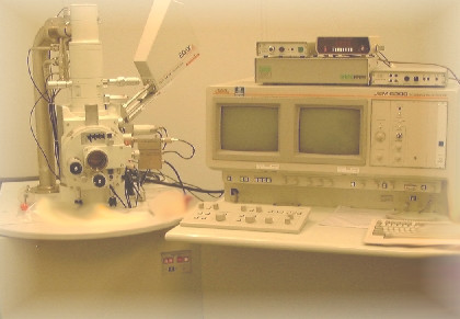 |
Joel JSM 6200 with Edax energy scattering x-ray analysis system. |
A scanning electron microscopy (SEM) is used to study samples in more detail than is possible using standard optical microscopy, for example foraminifera and mineral fragments, and to make chemical analyses from polished rock sections. SEM and x-ray analysis allows quantitative analysis of chemical elements in different parts of a mineral grain. Such variations can be used for geothermobarometry to determine the pressure and temperature at which the rock was formed or metamorphosed.
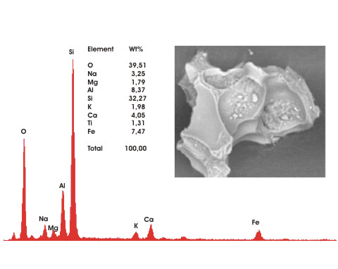 |
| Picture of an ash grain found in a sample from Andfjorden. The chemical composition, shown by the graph and table, indicates that the grain derives from a volcanic eruption on Iceland. The eruption occurred about 12 000 years ago. |
The Department of Geology shares the SEM with the Department of Medical Biology.
|


