|
Posisjon
1
Primary specificity of serine proteinases
|
Structural basis for
the primary specificity of serine proteinases of the chymotrypsin
family
The factors which determine
protein interaction and substrate specificity are poorly understood
because opposing energies are involved: The interactions are entropically
unfavourable due to e.g. reduced flexibility, but they can be associated
with gain in enthalpy due to burial of e.g. hydrophobic surfaces, or
formation of favourable electrostatic interactions.
Knowledge of
protein structure and ligand association is important in several fields,
such as:
- Diagnostics of diseases
- Approaches to drug design
-
Development of biological catalysts for industrial purposes
- Protein
design
Structure studies of protein complexes, in with
combination experimental biochemical data, mutational studies and
molecular dynamics simulations, may provide valuable information about the
factors influencing the function of the protein. Such knowledge is of
importance when trying to design proteins with highly specific functions.
The biophysical properties one wants to change can be:
- Increased efficiency
- Increased or decreased stability vs.
temperature, pH, salt etc.
- Increased or decreased specificity
Trypsin and chymotrypsin catalyse the hydrolysation of peptide
bonds. In order to compare the specificity of the two enzymes, the primary
binding residue of Bovine Pancreatic Trypsin Inhibitor (BPTI), lysine15,
was mutated to the other naturally occurring amino acids (except proline).
BPTI mimics a natural substrate, but in contrast to a normal catalysis,
the product is not released from the enzyme after hydrolysis.
|
|
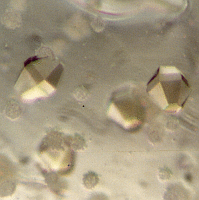
Orthorhombic crystals
of
Trypsin and BPTI
|
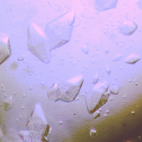
Hexagonal crystals of
Chymotrypsin and BPTI
|
The
association energies between the BPTI and bovine trypsin and chymotrypsin
have been measured, and it was found that the primary binding residue is
responsible for ca 70% of the binding energy in trypsin, and ca 50% in
chymotrypsin.
The 3-dimensional crystal structures of 10 variants
of BPTI in complex with trypsin and chymotrypsin have been determined. The
crystal structures show that the secondary binding interactions are almost
completely conserved in all complexes, and the P1 residues of the
inhibitor occupy almost identical conformations in the complexes with the
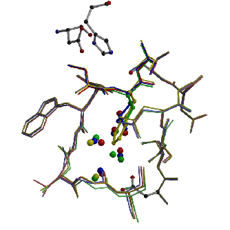
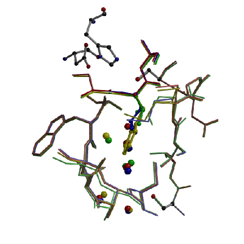
two enzymes. Three amino acids in the primary binding pocket of the two
enzymes differ, and this results in slightly different fold and polarity
of the pockets. The perturbation of the solvent network in the pocket and
unequal conformational strain imposed on the primary binding residues by
the pocket walls are the main cause of the differences in binding energies
and the different discrimination between primary residues of various size
and polarity.
|
|
Active site of trypsin with
4 mutants of BPTI:
Red:
glycine
Green: valine
Blue: leucine
Yellow:
Phenylalanine
|
Active site of chymotrypsin
with
4 mutants of BPTI:
Red:
glycine
Green: valine
Blue: leucine
Yellow:
Phenylalanine
|
References:
Krowarsch
et al. 1999.
J Mol Biol 289(1), 175-186.
Helland et al. 1999.
J. Mol. Biol. 287, 923 - 942.
Helland et al. 2003. J. Mol. Biol. 333, 845 - 861.
Czapinska et al. 2004 J. Mol. Biol. 344, 1005-1020.
|
|


