|
Atypical PKCs and p62 in Cellular Signaling
- Professor Terje Johansen
- Associate Professor Geir Bjørkøy
- Postdoctoral Researcher Trond Lamark
- Technician Aud Øvervatn
- Post doc Heidi Outzen
- PhD student Mikhail Sovershaev
- PhD student Serhiy Pankiv
- Medical student Terje Høgvarde Clausen
- PhD student Endalkachew Alemu
- Master student Camilla Sletten
- Master student Maria Jæger
Project description Publications Links
Project description
 |
|
Polypeptide-derived growth factors and activation of Ras or Src cause an increase in the hydrolysis of phosphatidylcholine (PC) resulting in a sustained elevation in intracellular levels of PC-derived diacylglycerol (DAG). Chronic stimulation of PC hydrolysis by stable expression of the gene encoding Bacillus cereus phosphatidylcholine-hydrolyzing phospholipase C (PC-PLC) causes severe growth deregulation and morphological transformation of NIH 3T3 fibroblasts without activating Ras. The generation of DAG by PC hydrolysis is located downstream of Ras but upstream of Raf in the mitogenic signal transduction pathway. The protein kinase C (PKC) family of lipid-dependent serine/threonine kinases plays pivotal roles in a wide variety of cellular processes. Based on sequence homology, domain organization and biochemical properties ten different isoforms are grouped into three classes denoted classical-, novel- and atypical PKCs (see Fig. 1). zPKC and l/iPKC constitute the atypical PKCs (aPKCs) .
 |
| Fig. 1. The PKC family of lipid-dependent protein kinases. |
While the classical and novel PKCs contain two repeated diacylglycerol (DAG)-binding zinc finger domains within their regulatory domains, the aPKCs have only a single zinc finger domain that is unable to interact with DAG or phorbol esters. Instead, atypical PKCs have been shown to be regulated in vitro and in vivo by other lipid products such as ceramide and phosphatidylinositol 3,4,5-trisphosphate, a product of phosphatidylinositol 3-kinase (PI 3-kinase). Consistently, aPKCs are strongly implicated as downstream effectors of PI 3-kinase. Recently, evidence has accumulated that imply important roles for aPKCs in processes as diverse as proliferation, cell survival, differentiation, cell polarity, insulin-mediated upregulation of general protein synthesis and glucose transport. Somewhat surprisingly, our work on Ras and PC-PLC transformed NIH 3T3 fibroblasts showed that the classical and novel PKCs were not essential for the mitogenic response observed upon a constitutive increase in the level of PC-derived DAG. However, we found that the transformed phenotype of v-Ras- or Bacillus cereus PC-PLC-expressing NIH 3T3 cells reverted upon expressing a kinase-defective mutant of lPKC. Extracellular signal-regulated kinase (ERK)-1 and -2 are constitutively activated in v-Ras- and PC-PLC-transformed cells in the absence of added growth factors. Interestingly, the activated ERKs are exclusively localized to the cell nucleus. Reversion of v-Ras- or PC-PLC-induced transformation by expression of dominant negative lPKC abolishes the nuclear ERK activation suggesting lPKC as a novel, direct or indirect, activator of mitogen-activated protein kinase/ERK kinase in response to activated Ras or elevated levels of PC-derived DAG. Transient transfection experiments confirmed that PKC acts downstream of Ras but upstream of mitogen-activated protein kinase/ERK kinase. Thus, when DAG cannot interact directly with lPKC how is this kinase activated by an elevation of the DAG level? It is possible that this occurs via ceramide which is increased following elevation of PC-derived DAG.
Our present focus is on how substrate specificity is achieved via anchoring or scaffolding proteins for aPKCs directing the kinases to different signalling complexes at different subcelleular localizations. In the course of these studies we have mapped a functional nuclear localization signal (NLS) to the N-terminal part of the zinc finger domain of lPKC. Leptomycin B treatment induced rapid nuclear accumulation of GFP-lPKC as well as endogenous lPKC suggesting the existence of a CRM1-dependent nuclear export signal (NES). Consequently, we identified a functional leucine-rich NES in the linker region between the zinc finger and the catalytic domain of . PKC. The presence of both the NLS and NES enables a continuous shuttling of lPKC between the cytoplasm and nucleus (Fig.2).
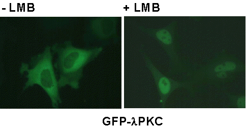 |
| Fig. 2. Treatment with LMB leads to rapid nuclear accumulation of a GFP-lPKC fusion protein. |

Publications
Johansen, T., Bjørkøy, G., Øvervatn, A., Diaz-Meco, T., Traavik, T. and Moscat, J. 1994. NIH 3T3 cells stably transfected with the gene encoding phosphatidylcholine-hydrolysing phospholipase C from Bacillus cereus acquire a transformed phenotype. Molecular and Cellular Biology, 14, 646-654.
Bjørkøy, G., Øvervatn, A., Diaz-Meco, T., Moscat, J. and Johansen, T. 1995. Evidence for a bifurcation of the mitogenic signalling pathway activated by Ras and phosphatidylcholine-hydrolyzing phospholipase C. Journal of Biological Chemistry, 270, 21299-21306.
Bjørkøy, G., Perander, M., Øvervatn, A. and Johansen, T. 1997. Reversion of Ras- and phosphatidylcholine-hydrolyzing phospholipase C-mediated transformation of NIH 3T3 cells by a dominant interfering mutant of lPKC is accompanied by the loss of constitutive nuclear mitogen-activated protein kinase/extracellular signal-regulated kinase activity. Journal of Biological Chemistry, 272, 11557-11565.
Perander, M., Bjørkøy, G. and Johansen, T. 2001. Nuclear import and export signals enable rapid nucleocytoplasmic shuttling of the atypical protein kinase C l. Journal of Biological Chemistry, 276, 13015-13024.
Lamark, T., Perander, M., Outzen, H., Kristiansen, K., Øvervatn, A., Michaelsen, E., Bjørkøy, G. and Johansen, T. 2003. Interaction Codes within the Family of Mammalian Phox and Bem1p Domain-containing Proteins. Journal of Biological Chemistry, 278, 34568-34581.
Bjørkøy, G., Lamark, T., Brech, A., Outzen, H., Perander, M., Øvervatn, A., Stenmark, H. and Johansen, T. 2005. p62/SQSTM1 forms protein aggregates degraded by autophagy and has a protective effect on huntingtin-induced cell death. J. Cell Biol. 2005 171, 603-614.
Links
- Protein Kinase Resource
- Scansite
- PhosphoBase
- Phosphoprotein Database Site
- Signal Transduction Knowledge Environment
- TransPath
- Biocarta
- Rac page
- Pawson Lab
- GeneCard for iPKC
- GeneCard for zPKC
- GeneLynx for iPKC
- GeneLynx for zPKC
- aPKC-review (J. Moscat)
- The extended PKC family (Mellor and Parker)
- Signalling through PKC - A. Toker
- Review on iPKC - Ohno
- Review on zPKC- Hirai and Chida
- Search PubMed for aPKC
|


